Remise du Prix scientifique de 500 000 euros de la Fondation Lefoulon-Delalande à l'institut de France.
Christine EDRY SEIDMAN
&
Jonathan G. SEIDMAN
LES GRANDS PRIX DES FONDATIONS DE L'INSTITUT DE FRANCE ONT ÉTÉ REMIS LE MERCREDI 13 JUIN 2007 SOUS LA COUPOLE:
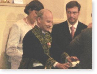
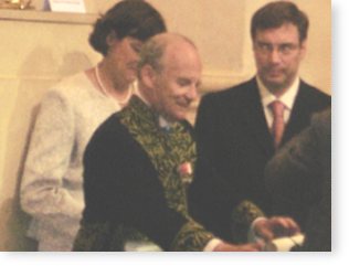
Le Prix scientifique de 500 000 euros de la Fondation Lefoulon-Delalande est remis par M. Alain CARPENTIER, de lAcadémie des sciences, aux professeurs : Christine EDRY SEIDMAN et Jonathan G. SEIDMAN du département Médecine et Génétique de la Harvard Medical School, de Boston, pour leurs travaux de recherche dans le domaine cardio-vasculaire.
Ce Prix a été remis lors de la Remise solennelle
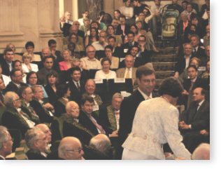
des Grands Prix des Fondations de lInstitut de France le mercredi 13 Juin 2007, à 15 heures sous la Coupole de lInstitut.
Ouverture par Madame Hélène CARRÈRE dENCAUSSE, Président de lInstitut de France Secrétaire perpétuel de lAcadémie française Introduction par Monsieur Gabriel de BROGLIE
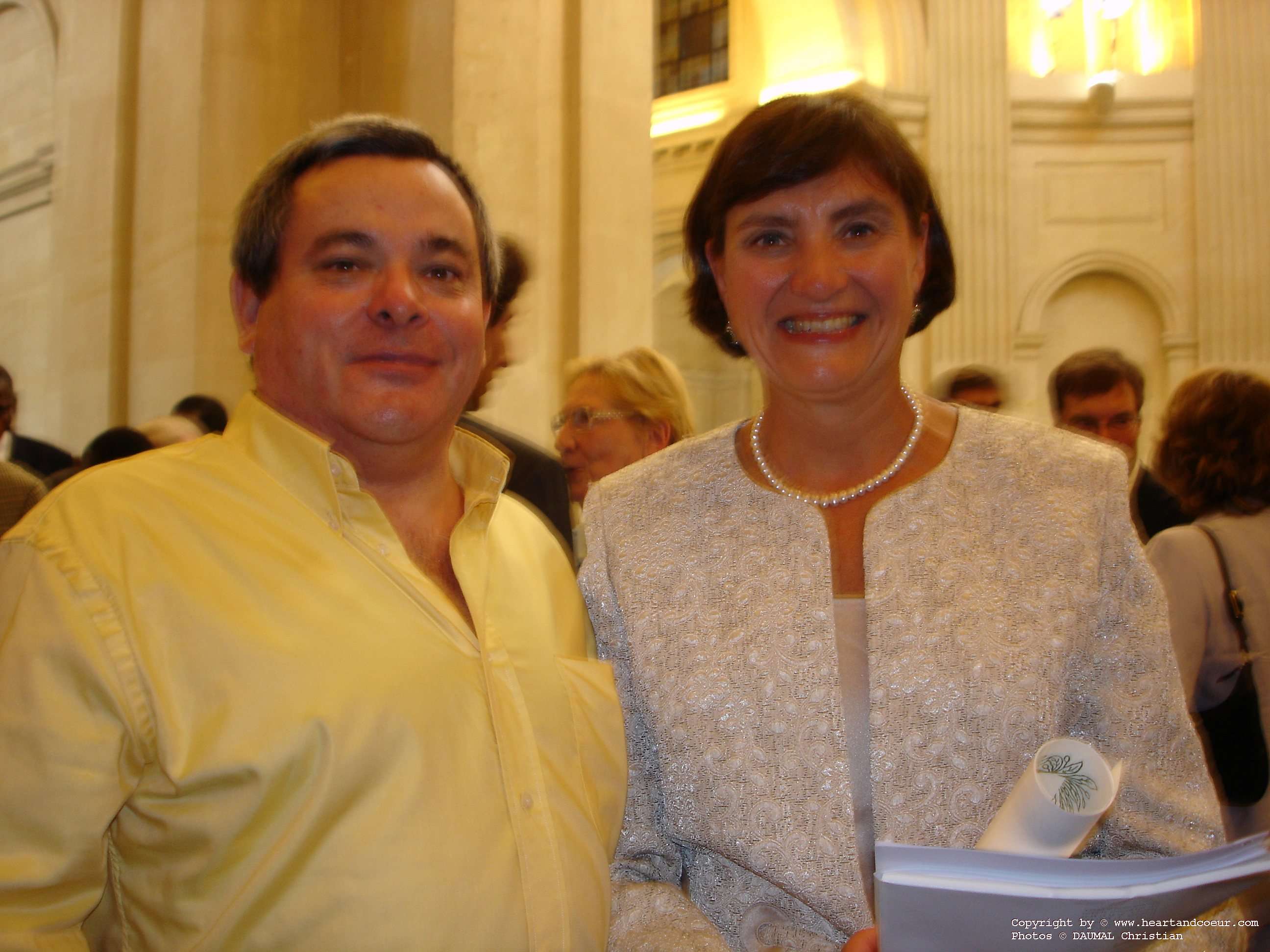
Chancelier de lInstitut L'association HEART and COEUR y était chaleureusement invitée pour représenter la cardiologie et les cardiopathies congénitales, et c'est avec honneur que Monsieur DAUMAL christian président de l'association, assistait à la remise du Prix scientifique de 500 000 euros de la Fondation Lefoulon-Delalande aux professeurs :
Christine EDRY SEIDMAN et Jonathan G. SEIDMAN du département Médecine et Génétique de la Harvard Medical School, Boston, pour leurs travaux de recherche dans le domaine cardio-vasculaire.
Résumé des recherches de Christine EDRY SEIDMAN et Jonathan G. SEIDMAN
Les docteurs Jonathan et Christine Seidman se sont distingués par leurs contributions fondamentales à la compréhension des bases moléculaires des maladies cardiaques humaines.
Ils ont découvert les mutations génétiques responsables des cardiomyopathies hypertrophique, métabolique et dilatée et des maladies congénitales du coeur.
En construisant des mutations humaines dans des organismes modèles, les Seidman ont déterminé des signaux pathogéniques importants pour ces maladies.
Leur travail améliore le diagnostic des maladies cardiaques, fournit des connaissances mécanistiques et permet de nouvelles opportunités de traItement.
Les études génétiques des Seidman ont commencé avec la cardiomyopathie hypertrophique (CMH), une maladie énigmatique caractérisée par une croissance inexpliquée du muscle cardiaque qui survient chez un individu sur cinq cents et explique la mort par arrêt cardiaque chez les athlètes.
En 1989, ils ont cartographié le premier gène de la CMH et ont ensuite mis en évidence les mutations responsables de la maladie dans le gène de la chaîne lourde bêta de la myosine cardiaque.
Cette découverte a conduit les Seidman à identifier d'autres gènes humains de la CMH, comme la troponine T cardiaque, l'alpha tropomyosine, la protéine - C de liaison de la myosine et la chaîne lourde alpha de la myosine cardiaque.
Collectivement, leur travail a établi la CMH comme une maladie du sarcomère.
En reproduisant des mutations du sarcomère humain chez la souris, les Seidman ont découvert des aspects inattendus de la physiopathologie de la CMH.
Ils ont trouvé que les mutations CMH dans la myosine augmentaient les fonctions biophysiques et ne les diminuaient pas, un résultat qui allait à l'encontre de l'idée classique selon laquelle l'hypertrophie était une réponse compensatoire à une fonction contractile réduite.
Les Seidman ont aussi découvert les causes génétiques des formes atypiques d'hypertrophie cardiaque, telles que des mutations dans la sous-unité gamma-2 régulatrice de la protéine kinase activée par l'AMP (PRKAG2) et dans la protéine de membrane 2 associée au lysosome (MP-2).
Leurs études ont révélé des manifestations physioopathologiques distinctes par lesquelles ces mutations provoquaient l'hypertrophie qui a expliqué les arythmies accompagnant ces défauts génétiques et mettant en jeu le pronostic vital.
Les causes génétiques de l'insuffisance cardiaque ont été largement étudiées par les Seidman. Ils ont identifié des mutations chez l'homme dans la lamine A, protéine d'enveloppe nucléaire, la cause génétique la plus courante de cardiomyopathie dilatée accompagnée d'anomalies électrophysiologiques, et dans le phosspholamban, qui perturbe la recapture du calcium dans le réticulum sarcoplasmique.
Les Seidman ont aussi décrit des mutations dans les gènes des protéines du sarcomère et ont montré qu'elles avaient des conséquences réciproques sur les fonctions biophysiques modifiées par les mutations hypertrophiques.
Les études des Seidman sur les causes génétiques de la maladie congénitale du coeur a conduit à la découverte de mutations dans les gènes des facteurs de transcription NKX2.5 et TBX5.
Leur travail a établi le paradigme que des taux inadaptés de facteurs de transcription cardiaques pendant le développement sont la cause de la survenue de malformations cardiaques.
Leur groupe a produit des modèles de maladie cardiaque congénitale et a défini les cibles en aval des facteurs de transcription qui sont critiques pour la morphogenèse cardiaque, la septation et la formation du système électrophysiologique cardiaque.
Les études génétiques cardiovasculaires des Seidman permettent le diagnostic moderne, basé sur les gènes, de plusieurs maladies humaines, elles ont fourni un cadre moléculaire à la compréhension de situations cardiaques complexes et ont montré le bénéfice de l'information génotypique pour l'amélioration de la prise en charge des malades.
Un lieu prestigieux pour la remise des prix: l'INSTITUT de FRANCE.
L'Institut de France regroupe cinq académies. L'habit vert, la cape, le bicorne et l'épée, portés par tous les académiciens membres de l'Institut de France lors des séances solennelles sous la Coupole, remontent au Consulat.
Parlement du monde savant, il a pour missions :
de perfectionner les arts et les sciences selon le principe de pluridisciplinarité ;
de gérer le millier de dons, legs et fondations dont il est dépositaire, se donnant ainsi les moyens dexercer la première de ses missions.
Il est le regroupement de cinq académies :
lAcadémie française (fondée en 1635)
lAcadémie des inscriptions et belles-lettres (fondée en 1663)
lAcadémie des sciences (fondée en 1666)
lAcadémie des beaux-arts (créée en 1816 par la réunion de lAcadémie de peinture et de sculpture, fondée en 1648, de lAcadémie de musique, fondée en 1669 et de lAcadémie darchitecture, fondée en 1671)
lAcadémie des sciences morales et politiques (fondée en 1795, supprimée en 1803 et rétablie en 1832).
L'Institut comprend aussi un service des actions pédagogiques et dispose d'un service de publications.
Metabolic Causes of Cardiac Hypertrophy ( English version)
Metabolic Causes of Cardiac Hypertrophy
Summary: Christine Seidman's laboratory discovers gene mutations that cause human disease, with a focus on cardiovascular conditions such as cardiomyopathy (hypertrophic and dilated), heart failure, and congenital heart malformations. Her laboratory also produces model organisms that carry human mutations, and uses these models to determine how responses to gene mutations perturb or influence myocardial structure and specialized heart functions.
The development of left ventricular hypertrophy (LVH) is an adverse and independent risk factor for a cardiovascular event and an important contributing factor in the development of heart failure. LVH is a common response by the heart to a variety of stressful stimuli, including increased blood pressure, elevated body mass index, insulin resistance, myocardial infarction, and valvular heart disease. Unexplained‿ LVH, with an absence of known triggers for hypertrophic remodeling, is often a primary myocardial disease caused by a gene mutation.
To study the signaling pathways that produce LVH, we have defined human mutations that cause cardiac hypertrophy. Our initial investigations focused on hypertrophic cardiomyopathy (HCM), an autosomal-dominant disorder characterized by unexplained hypertrophy and histopathologic findings of myocyte and myofibrillar disarray. Mutations in 10 different sarcomere protein genes are now known to cause HCM. Sarcomere protein gene mutations also cause cardiac hypertrophy that is not readily recognized as a heritable trait, such as occurs in sporadic HCM or elderly onset HCM. To discover other genetic causes of LVH, we have focused on subjects with a clinical diagnosis of HCM but in whom a sarcomere gene mutation was not identified.
Clinical evaluations of our subjects indicated two unique cohorts. One group had LVH that was inherited as a dominant trait, but unlike subjects with sarcomere gene mutations, this cohort often had cardiac electrical abnormalities. Electrophysiologic manifestations included ventricular pre-excitation (indicating an abnormal pathway for impulse propagation from the atria to ventricles) atrial fibrillation, and over time, progressive deterioration in impulse propagation, such that pacemaker implantation was required to maintain cardiac conduction. We recently defined the genetic cause of this cardiomyopathy to be mutation in the γ2 subunit of AMP-dependent protein kinase (AMPK). AMPKa heterotrimeric protein consisting of α, β, and γ subunits that detect cellular changes and modify substrate utilization for energy production. AMPK binds glycogen, phosphorylates proteins, and modifies gene expression.
To understand the mechanisms by which mutations in the γ2 subunit (denoted PRKAG2) cause cardiac hypertrophy, we engineered human mutations into the yeast homolog of γ2 subunit and examined the biochemical interactions among AMPK subunits. In the absence of glucose, yeast homologs of the α and γ subunits interact, but when glucose is present, this association is normally inhibited. However, when a yeast homolog of γ2 subunit carried a human PRKAG2 mutation, subunit interactions were not inhibited by glucose. This suggested that human PRKAG2 mutations result in constitutive AMPK activity, a finding that implies abnormal glycogen metabolism and glycogen storage as one mechanism for cardiac hypertrophy. Cardiac histopathology of specimens from subjects with PRKAG2 mutations support this model. Unlike HCM hearts, PRKAG2 mutations cause myocyte hypertrophy and myocyte vacuoles that are filled with glycogen and amylopectin, an insoluble remnant of glycogen. The cellular events triggered by constitutively active PRKAG2 result in cardiac glycogen storage and conduction system disease, which we are modeling in mice.
We also identified a second cohort of patients with LVH that mimicks HCM. Like patients with PRKAG2 mutations, this cohort had electrophysiologic abnormalities. More often, however, these patients had massive LVH, with a ventricular wall thickness that sometimes exceeded 50 mm. (Sarcomere protein gene mutations and PRKAG2 mutations typically cause more modest hypertrophy, with wall thickness usually less than 25 mm. The normal ventricular wall is 1315 mm). Other clinical features that distinguish this cohort include male gender, childhood onset of disease, progression to heart failure in young adulthood, and the absence of a family history of cardiomyopathy.
Based on the clinical findings associated with PRKAG2 mutations, we hypothesized that mutations in other cardiac glycogen metabolism genes might account for massive LVH and electrophysiologic abnormalities. We performed direct DNA sequence analyses of candidate genes to test this model. Our analyses of the X-linked gene encoding lysosome-associated membrane protein (LAMP2) found multiple independent mutations. More than half of these are de novo mutations. LAMP2 encodes a 410amino acid molecule with a small cytoplasmic tail involved in receptor-mediated uptake and a large internal lysosome domain composed of highly glycosylated residues. Several mutations encode a truncated peptide; others encode altered splice signals. To assess whether mutant LAMP2 was produced, we performed RNA and protein studies of lymphocyte and cardiac or skeletal muscle specimens. Surprisingly, both stable mutant LAMP2 RNA and immunoreactive LAMP2 protein were found in some specimens.
LAMP2 mutations have been previously demonstrated to cause Danon disease, a systemic disorder characterized by mental retardation, musculoskeletal weakness, and hepatic disease. No subjects in our series had clinically recognized neurological disease nor overt muscle weakness, wasting, or myopathic symptoms. The identification of LAMP2 mutations prompted retrospective analyses of serum chemistries; two-thirds of the subjects had unrecognized mild elevations of cardiac, skeletal muscle, and liver isoforms of creatine phosphokinase and serum pyruvic glutamic transaminase levels.
Two mechanisms might account for the cardiac-restricted phenotype observed with LAMP2 mutations that cause primarily LVH versus other mutations that produce the pleiomorphic findings of Danon disease. First, some mutant LAMP2 protein is stably produced in cells (as evidenced by immunohistochemistry) and may retain sufficient partial function so as to limit disease in some organs, although not in the heart. Gene dosage also appears to play a role in the clinical expression of LAMP2 mutations. We found an identical LAMP2 mutation in two unrelated male subjects; genetic mosaicism appears to have conferred cardiac-restricted disease in one, while the other individual was hemizygous for the LAMP2 mutation and had classic Danon disease. A LAMP2 mutation was also found in a woman with profound cardiac hypertrophy, implying that X inactivation may have sufficiently extinguished normal LAMP2 gene expression to contribute to or cause her cardiomyopathy.
PRKAG2 and LAMP2 mutations define a new mechanism for cardiac hypertrophypathologic glycogen metabolism by the heart. Although LAMP2 mutations accumulate glycogen in lysosomes and PRKAG2 mutations accumulate glycogen throughout the myocyte, we suspect these defects trigger a common pathway that remodels the ventricle and damages the cardiac conduction system. Understanding these mechanisms may provide opportunities to treat these cardiac storage diseases. Even before then, recognition of different genetic etiologies for cardiac hypertrophy has clinical value. The profoundly different clinical courses and management issues associated with HCM or glycogen storage cardiomyopathy underscore the importance of accurate diagnosis. Although the correct diagnosis can be suggested from subtle differences in clinical manifestations, gene-based diagnosis accurately defines the molecular causes of unexplained cardiac hypertrophy.
Partial support for these studies comes from grants from the National Institutes of Health. http://www.hhmi.org
Publications
1989 Jarcho J-J., McKenna W]., Pare JAP., Solo mon S.D., Holcombe R.F., Dickie S., Levi T., Seidman ].G., Seidman CE. "Mapping a Gene for Familial Hyperrrophic Cardiomyopathy to Chromosome 14ql", N Eng.j. Med., 321(20) :1372-8.
1990 Geisterfer-Lowrance AAT., Kass S., Tanigawa G., Vosberg H.P., McKenna, Seidman J.G. and Seidman CE. "A Molecular Basis for Familial Hypertrophie Cardiomyopathy : A fi Cardiac Myosin Heavy Chain Gene Missense Mutation", Cell 62: 999-1006.
1991 Rosenzweig A, Watkins H., Hwang D-S., Miri M., McKenna W, Trail! TA, Seidman J.G., Seidman CE. "Hematologie identification of myosin mutations permits pre-clinical diagnosis of Familial Hypertrophie Cardiomyopathy", N Eng.j. Med., 325(25): 1753-60 .
1992 Watkins H., RosenzweigA, Hwang D-5, Levi T., McKenna W, Seidman CE., Seidman J.G. "Distribution and clinical implications of myosin missense mutations that cause familial hyperrrophic cardiomyopaathy", N Eng.j. Med., 326(17): 1108-14 .
1994 Basson CT., Cowley G.S., Solomon S., Weissman B., Stern A, Poznanski AK., Traill T.A., Seidman ].G. and Seidman CE. "The cliniical and genetic spectrum of Holt-Oram Syndrome", N Eng. j. Med., 330 : 885-891.
Thierfe!der L., Watkins H., MacRae C, Lamas R., McKenna W, Vosberg H-P., Seidman ].G. and Seidman CE. "A-Tropomyosin and carrdiac troponin T mutations cause familial hypertrophie cardiomyopathy: A disease of the sarcomere", Cell 77 : 1-20.
1996 Geisterfer-Lowrance AAT., Christe M., Conner D.A, Ingwall ].S., Schoen F.]., Seidman CE., Seidman J.G. "A murine mode! of famillial hypertrophie cardiomyopathy", Science, 272: 731-734.
1997 Basson CT., Bachinsky D.R., Lin R.C, Levi T., Elkins JA, Soults]., Grayze! D., Kroumpouzou E., Traill T.A., Leblanc-Straceski J., Renault B., Kucherlapati R., Seidman J.G., Seidman CE. "Mutations in human TBX5 cause limb and cardiac malformation in Hoit-Oram synndrome", Nat Genet, 15 : 30-35.
1998 Schott,]']" Benson D.W, Basson CT., Pease W, Silberbach M., Moak J.P., Maron B.J., Seidman CE., and Seidman ].G., "Congenital Heart Disease caused by Mutations in the Transcription Factor Nkx2.5", Science, 281 : 108-111.
1999 Fatkin D., MacRae C, 5asaki T., Wolff M.R., Porcu M., Frenneaux M., Atherton ]., Vidaillet H.J., Spudich S., DeGirolami D., Muntoni F., Johnson W., McDonough B., Seidman J.G., Seidman CE., "Missense mutations in the lamin NC tod cause dilated cardiomyopathy and conduction system disease", N Eng. j. Med., 341(23) :1715-1724.
2001 Bruneau BG, Nemer G, Schmitt JP, Charron F, Robitaille L, Caron S, Conner DA, Gessler M, Nemer M, Seidman CE, Seidman JG., "A Murine Mode! of Holt-Oram Syndrome Defines RoIes of the T-Box Transcription Factor Tbx5 in Cardiogenesis and Disease", Celll06: 7099721.
2003 Schmitt]" Kamisago M., Asahi M., Li G.H., Ahmad F., Mende D., Kranias E.G., MacLennan D.H., Seidman J.G. and Seidman, CE. "Inactivation of Protein Kinase A by Phospholamban Arg9Cys Causes Dilated Cardiomyopathy and Heart Failure in Mice and Man", Science, 299 : 1410-1413.
2005 Arad M, Maron BJ, Gorham JM, Johnson WH, Saul JP, PerezzAtayde AR, Spirito P, Wright, GB, Kanter RJ, 5eidman CE and Seidman JG., "Glycogen storage diseases presenting as hypertrophie cardiomyopaarhy". N Engl. j. Med., 352(4) : 362-372.
2007 Kim J.B., Potteea G., Song L., Greenway S., Gorham J.M., Church G.M., Seidman CE., Seidman ].G. "Polony multiplex analysis of gene expression (PMAGE) of the adult mouse heart", Science, in press.